Große Auswahl an günstigen Büchern
Schnelle Lieferung per Post und DHL

Bücher veröffentlicht von Thieme Medical Publishers

Filter
Filter
Ordnen nachSortieren Beliebt
  • von Jörg Kastner
    73,00 €

    Let food be your medicine and medicine be your food – Hippocrates, ca. 460–ca. 370 BCFor millennia, the Chinese have taught that a balanced and nutritious diet is an integral part of maintaining good health while healing a wide range of disorders. Here, in a new and third edition of the acclaimed Chinese Nutrition Therapy, the author further demystifies Chinese dietetics, one of the pillars of Traditional Chinese Medicine (TCM).Joerg Kastner's introduction to the principles of TCM and the "energetics" of foods emphasizes a holistic approach throughout, providing readers with tools for integrating the principles of Chinese dietetics into their daily lives. Included are a comprehensive classification of more than 300 readily available foods such as vegetables, grains, herbs and spices, fruits, meat and poultry, seafoods, dairy products, oils and fats, and nuts and seeds. Many clinical examples and treatment suggestions artfully combine Western medical conditions with Chinese diagnostic and therapeutic techniques.

  • von Torsten Möller
    44,98 €

    This comprehensive, easy-to-consult pocket atlas is renowned for its superb illustrations and ability to depict sectional anatomy in every plane. Together with its two companion volumes, it provides a highly specialized navigational tool for all clinicians who need to master radiologic anatomy and accurately interpret CT and MR images. Special features of Pocket Atlas of Sectional Anatomy: Didactic organization in two-page units, with high-quality radiographs on one side and brilliant, full-color diagrams on the other Hundreds of high-resolution CT and MR images made with the latest generation of scanners (e.g., 3T MRI, 64-slice CT) Consistent color coding, making it easy to identify similar structures across several slices Concise, easy-to-read labeling of all figures Updates for the 4th edition of Volume I: New cranial CT imaging sequences of the axial and coronal temporal bone Expanded MR section, with all new 3T MR images of the temporal lobe and hippocampus, basilar artery, cranial nerves, cavernous sinus, and more New arterial MR angiography sequences of the neck and additional larynx images Compact, easy-to-use, highly visual, and designed for quick recall, this book is ideal for use in both the clinical and study settings.

  • von Andre Braz
    177,00 €

    In-depth and detailed knowledge of anatomy, different facial shapes, and ethnic origins is essential to delivering safe, effective, natural, and harmonious aesthetic facial treatment results. Dermal Fillers: Facial Anatomy and Injection Techniques is the culmination of many years of expertise decrypting facial anatomy as it applies to aesthetic enhancement of the face. This visually rich atlas is authored by internationally renowned Brazilian dermatologists André Braz, an esteemed expert in the use of botulinum toxin, fillers, and lasers; and Thais Sakuma, a distinguished lecturer in the application of botulinum toxin and hyaluronic acid fillers.Nineteen highly practical, concise, and didactic chapters are organized consistently with structured text at the beginning of each chapter. The book encompasses the application of injection techniques for the full spectrum of indications, from softening wrinkles to facial contouring. Important topics include facial assessment, hyaluronic acid science, facial aging, and regional nerve blocks for pain management. Meticulous anatomic descriptions and images depict the relationships between superficial and deeper structures such as vasculature, thereby providing vital knowledge to help clinicians avoid potentially serious complications.

  • von Stephen Thomas
    65,00 €

    RadCases Gastrointestinal Imaging Second Edition by Stephen Thomas and Jonathan Lorenz expands on the rich study experience that has been tried, tested, and popularized by radiology residents around the world. The new edition includes important variations on prior cases, updated diagnostic and management strategies, and new pathological entities. One hundred new, carefully selected GI cases are focused on helping radiology residents navigate and assimilate a daunting volume of digestive-related information. For maximum ease of self-assessment, each case begins with the clinical presentation on the right-hand page; study that and then turn the page for imaging findings, differential diagnoses with the definitive diagnosis, essential facts, pearls and pitfalls, and more.

  • von Z. Gromley
    82,00 €

    Biochemistry, Cell and Molecular Biology, and Genetics: An Integrated Textbook by Zeynep Gromley and Adam Gromley is the first to cover molecular biology, cell biology, biochemistry (metabolism), and genetics in one comprehensive yet concise resource. Throughout the book, these topics are linked to other basic medical sciences, such as pharmacology, physiology, pathology, immunology, microbiology, and histology, for a truly integrated approach.

  • von A Primer
    168,00 €

    Minimally Invasive Spine Surgery: A Primer by Luis Manuel Tumialán is the ideal introduction to minimally invasive spine approaches, especially for neurosurgery and orthopedic residents, fellows, and spine surgeons who want to incorporate minimally invasive approaches into their practice. The Primer offers a treasure trove of 3D illustrations and animations that virtually brings the aspiring minimally invasive spine surgeon into the operating room alongside their professor. The text starts with a discussion of open spine surgery versus minimally invasive procedures and the optimal mindset required to convert from one to the other.The book is divided into lumbar, cervical, and thoracic spine sections, and a fourth section dedicated to the fundamentals of fluoroscopy and radiation exposure. The text begins with an overview, history, and evolution of each procedure, followed by a discussion of the anatomical basis for using a minimally invasive approach. Each anatomical section starts with the least complicated surgeries, thereby laying the foundation for more complex procedures discussed in subsequent chapters. The third section focuses on thoracic decompression, nerve sheath tumors in the lumbar and thoracic spine, and management of metastatic disease and intradural extramedullary lesions.

  • von Miquel Videla Ces
    155,00 €

    The surgical profession places high demands on surgeons who must often make the right split-second decisions. This can easily lead to misjudgments or mistakes.Learning From Failures in Orthopedic Trauma—Key Points for Success is the first book of its kind to give surgeons the opportunity to learn from failures without making them themselves.Based on the Spanish book Errores en la Osteosíntesis by Rafael Orozco Delclós, this publication offers real case examples that have been collected over the past 25 years. It is an essential and valuable resource as it specifically examines the reasons and responses to surgical error in real cases from different anatomical regions of the body, thus helping surgeons avoid the most frequent errors in osteosynthesis.

  • von Thomas Bohmeke
    49,00 €

    In diagnostic cardiology, the usefulness and effectiveness of state-of-the-art echocardiography is unsurpassed. This handy atlas includes all of the information you need to navigate the numerous imaging planes that transect the heart with ease and confidence. Comprehensive coverage: More than 400 illustrations, including, sharp, clear echocardiograms, full-color schematic diagrams, and 3-D images. Detailed descriptions of all of the acoustic windows and imaging planes for every echocardiogram. All major diseases depicted in B-mode, M-mode, Doppler and color Doppler. A practical overview of the patient examination, including imaging and patient positioning. All cardiac diseases are shown - valvular heart disease, coronary heart disease, cardiomyopathies, prosthetic valves, carditis, septal defects, hypertensive heart diseases, intracardiac masses. Hundreds of vivid mnemonic devices and useful tips to help you locate, name, and remember all anatomical structures and features. Intelligent design: Integrated illustrations and succinct text on every page. Fits in your pocket for rapid reference and review. Durably designed to withstand everyday use.

  • von Michael Schutz
    160,00 €

    Hear the authors discuss Periprosthetic Fracture Management. This textbook accumulates the latest global knowledge on periprosthetic fractures, including all relevant anatomical regions, surgical pitfalls, complex cases, and a brand new comprehensive Unified Classification System, (UCS) on periprosthetic fractures, combining the original Vancouver classification with the AO/OTA Fracture and Dislocation Classification. Carefully selected case studies illustrate and describe individual solutions for often problematic fracture situations, providing comprehensive information from experts globally. Orthopedic and trauma surgeons will be able to expand their knowledge regarding: Risk factors and assessment of periprosthetic fractures and the patient A new Unified Classification System (UCS) Options and choices of fracture fixations techniques as well as more complex revisions or reconstructive procedures More than 350 pages containing superb illustrations and images.

  • von Simone Waldt
    111,00 €

    For all radiology residents and practitioners faced with mastering the huge array of image-based measurements and classifications in orthopedics, Measurements and Classifications in Musculoskeletal Radiology fills an important gap.Highly practical and accessible, it presents the measurement techniques, classification systems, scoring methods, and reference values currently in use in musculoskeletal radiology in a single, comprehensive volume, stressing the significance of each for determining the stage, diagnosis, treatment, and prognosis of a wide range of orthopedic disorders. Special Features: Gathers all currently fragmented information on musculoskeletal radiology measurements and classification systems (excluding bone fractures) into one convenient text– eliminating the need for time-consuming memorization and literature searches Covers all established measurement methods made using conventional and sectional imaging techniques Includes nearly 400 detailed illustrations and radiological images showing reference lines, markings, orientations, and angles, with key values indicated throughout Logically structured by anatomic sites and Pathologys for easy access to information, with clinical pearls highlighted in every chapter Offers practical guidance on the relevance and reliability of each imaging method, preferred techniques for specific diseases, and tips for achieving the clearest and most precise measurement results Ideal for use at the radiology workstation, this generously illustrated resource will also be helpful to orthopedic specialists and trauma surgeons who utilize radiologic measurements and classifications for optimal diagnostic and treatment decisions.

  • von Daniel Simmen
    160,00 €

    [This book is]one of the most comprehensive surgical guides I’ve seen in my career. It is a strongly how-to and problem-oriented tome. It was well written and very easy to read. The format makes it is easy to fi nd answers and specific recommendations. I would strongly recommend that all endoscopic sinus surgeons review the book and see if they can benefi t from the incredible expertise represented in this manual. -- The American Journal of Cosmetic Surgery Illustrated with hundreds of outstanding endoscopic images and corresponding drawings, this successfully established step-by-step guide to endoscopic sinus and skull base surgery is ideal for all specialists who need to stay current in the field. In this expanded second edition, practitioners will not only find basic and advanced procedures in endoscopic sinus surgery (including key criteria for patient selection), but also four entirely new chapters on anterior skull base surgery that reflect important recent developments. Special features of the second edition: Superbly illustrated, presenting over 1400 endoscopic images, photographs, and drawings Covers the latest techniques for removal of skull base tumors and repair of skull base defects Includes new chapters on pathophysiology of rhinosinusitis, sense of smell, applied anatomy, the endoscopically assisted bimanual operating technique, transorbital surgery, interdisciplinary teamwork, and more Offers online access to 34 videos of endoscopic surgical procedures and patient interviews, for an excellent review and refresher Provides Information for Patients leaflets that can also be downloaded From diagnosis and preoperative counseling to evidence-based procedures, complication avoidance, and postoperative management, this book covers the gamut of topics clinicians face in everyday practice. All otolaryngologists, neurosurgeons, and maxillofacial surgeons will find it essential when making diagnostic and treatment decisions involving the paranasal sinuses.

  • - Principles, Indications, and Techniques
    von Daniele Marchioni
    164,00 €

    Increasingly used as an adjunctive tool in the diagnosis and management of ear disease, middle ear endoscopy has the potential to decrease patient morbidity, prevent disease recurrence, and reduce costs. Its capacity to uncover “hidden” anatomy within the small dimensions of the ear has made it essential for functional surgery, allowing surgeons to preserve key anatomic structures such as the cochlea and facial nerve. Now for the first time, the physicians who pioneered this groundbreaking minimally invasive technique review its indications, advantages, disadvantages, and surgical approaches: Written by the foremost leaders in the field, all members of the International Working Group on Endoscopic Ear Surgery Features nearly 1,000 intraoperative endoscopic images filmed with high-definition cameras, plus outstanding line drawings that aid in understanding all concepts Comprehensively covers the field, including chapters on endoscopic and combined endoscopic-microscopic management of cholesteatoma, surgical restoration of middle ear ventilation, myringoplasty, Meniere disease, tympanoplasty and stapes surgery, revision surgery, and much more Demonstrates state-of-the-art endoscopic procedures in 19 instructive videos From the unique anatomy and pathophysiology of the middle ear through diagnosis, surgical indications, procedures, and instrumentation, this book is indispensable for all otolaryngologists and neurotologists. It offers a full picture of the field today, as well as exploring the future possibilities of ear endoscopes alone or in combination with operative microscopes for the most effective treatment of middle ear Pathologys. Thieme eOtolaryngology is the premier online resource for otolaryngology-head and neck surgery.

  • von Martin Rocken
    44,98 €

    Color Atlas of Dermatology is an illustrated guide to the broad spectrum of dermatological pathologies. This easy-to-use book provides a wealth of practical knowledge essential in both daily practice and when planning treatment.The atlas organizes the main areas of clinical dermatology in a user-friendly formatpresenting short, precise descriptions on the left and illustrative images on the right. This text brings you the most practical and comprehensive information. It begins with the basic principles of dermatological practice, followed by a thorough study of each disease accompanied by techniques for diagnoses and treatment. Treatment procedures include surgery, pharmacotherapy, and the physical modalities. The instructive color plates are an invaluable aid in visualizing even the most complex relationships.Features:Hundreds of detailed images of actual cases, including graphic illustrations of diagnostic and therapeutic pathwaysValuable treatment notes for all common dermatological conditionsAll of the current scientific knowledge of dermatology combined with the classic Thieme Flexibook layout with a double-page spreadThe Color Atlas of Dermatology is a handy clinical reference for dermatologists and general practitioners, as well as an excellent review tool for medical students.

  • von Gert Krischak
    62,00 €

    This is a good book for clinicians looking for more information about the surgical management of fractures and, overall, it is a fine reference for practicing clinicians in acute care and orthopedics as well as ambitious students. -- Doody's Enterprises, Inc. For physical therapists working in hospitals, rehabilitation centers, and in private practice, knowledge of commonly seen traumatic and orthopedic injuries is essential to sound treatment planning. This book offers a focused, easy-to-use guide to general and specialized traumatology specifically geared to physical therapists and students. From types of injuries and resulting symptoms to diagnosis and treatment, the book brings a structure to treatment planning and greatly improves the communication between patient, physical therapist, and physician. Special Features: Covers all relevant medical information for physical therapists, including general traumatology (wound healing, surgical infections, soft tissue injuries, and fractures), special traumatology (cranial, spinal, thoracic, and abdominal injuries), and multiple trauma and first aid Offers guidelines on the benefits, effects, and limitations of physical therapy across a wide range of injuries and conditions Supplies 326 full-color illustrations that clarify every concept Includes bulleted summaries at the end of each chapter, for an instant review of the material Provides quiz questions and a glossary of medical terms at the end of each section Shares the unique perspective of a practicing physician who also teaches physical therapy at a major international rehabilitation center Providing a broad understanding of the pathologic basis of traumatic injuries and its effect on restoring function, this book is a key resource on formulating effective physical therapy strategies. Practicing physical therapists, occupational therapists, and physical therapy students will find it an invaluable learning and practice tool.

  • - Japanese Pediatric Acupuncture, A Text and Video Guide
    von Stephen Birch
    78,00 €

    Now in an expanded new edition, this user-friendly manual remains the foremost instructional reference for the application of Shonishin, or Japanese pediatric acupuncture. The book addresses the unique and often changing circumstances involved in the treatment of infants and children. It focuses on the crucial factors of patient comfort and dosage regulation throughout treatment by gently and painlessly pressing, tapping, scratching, rubbing, and stroking the skin without penetrating it. Stephen Birch, the world’s leading authority in Japanese acupuncture and Shonishin, provides valuable clinical guidance and hands-on advice that can be easily integrated into everyday practice. The book begins by presenting the underlying principles and treatment tools used in Shonishin, and then goes on to cover root and symptomatic approaches and techniques, followed by details on how to manage a wide range of specific problems and diseases. More than 75 case histories from around the world present various treatment ideas, methods, and results for the featured condition. Highlights: More than 25 new clinical cases (over 75 cases total) offer useful insights and suggestions for daily practice New information on using Shonishin to treat headaches and emotional problems; combining Shonishin with other treatments, such as Bach flowers and Chinese herbal medicine; and using the techniques of Shonishin and the Meridian Therapy root treatment Coverage of Shonishin for the management of a broad spectrum of illnesses and diseases, including respiratory and digestive ailments, ear infections, allergies, sleep problems, and many more Exercises throughout the book help readers improve their skill and check their technique More than 150 precise illustrations complement and further clarify the text Video footage of a workshop where the author demonstrates how to do Shonishin and shows real treatments bring concepts to life (accessed through Thieme’s Media Center).

  • - Thorax, Heart, Abdomen and Pelvis
    von Torsten Bert Moller
    44,98 €

    4th revised edition 2013. This comprehensive, easy-to-consult pocket atlas is renowned for its superb illustrations and ability to depict sectional anatomy in every plane. Together with Volumes I and III, it provides a highly specialized navigational tool for all clinicians who need to master radiologic anatomy and accurately interpret CT and MR images. Special features of Pocket Atlas of Sectional Anatomy: Didactic organization in two-page units, with high-quality radiographs on one side and brilliant, full-color diagrams on the other. Hundreds of high-resolution CT and MR images made with the latest generation of scanners (e.g., 3T MRI, 64-slice CT) Color-coded schematic drawings that indicate the level of each section. Concise, easy-to-read labeling of all figures. Sectional enlargements and magnified views for easy classification of anatomic structures Updates for the 4th edition of Volume II: CT imaging of the chest and abdomen in all 3 planes: axial, sagittal, and coronal. New sections on MRI and CT of the heart and MR angiography. New back-cover foldout featuring pulmonary and hepatic segments and lymph node stations. Follows standard international classifications of the American Heart Association for cardiac vessels and the AJCC/UICC for mediastinal lymph nodes. Compact, easy-to-use, highly visual, and designed for quick recall, this book is ideal for use in both the clinical and classroom settings.

  • von Jerome Lam
    115,00 €

    1. udgave. Mobile ultrasonography is revolutionizing the way hand surgery patients are managed. Ultrasound of the Hand and Upper Extremity: A Step-by-Step Guide is the first book on this field that is intended for practicing hand surgeons and the radiologists who work with them. A stepwise, practical guide specially designed for quick reference, with bullet-point text, informative figures, and detailed clinical examples, this book and its accompanying videos are ideal for the busy clinician. Edited by John R. Fowler and Nandkumar M. Rawool, with contributions by other experts with long experience in ultrasound techniques, this book features a reader-friendly chapter structure that describes the appropriate setup, anatomic landmarks, probe and patient positioning, comparative normal anatomy, relevant pathologic anatomy, and available injection techniques for 14 anatomic areas and conditions.

  • von Gillian M. Newstead
    135,00 €

    Magnetic resonance imaging (MRI) of the breast has evolved into an important breast cancer screeningtool and major advance in women's health. Breast MRI is currently the most sensitive detection techniquefor both non-invasive and invasive cancers and follow-up in women with a new breast cancer diagnosis. Itis increasingly becoming the go-to imaging method for screening women at high and intermediate risk ofbreast cancer and those with dense breast tissue on mammography. Yet despite its reliability andgrowing use, many radiologists lack the expertise to accurately perform breast MR image interpretation.Breast MRI Interpretation: Text and Online Case Analysis for Screening and Diagnosis by Gillian Newsteadreflects insights and expertise from one of the leading authorities on breast imaging. The book is a highlypractical reference on evaluation and interpretation of breast MR imaging, with discussion of the modalityas a screening and diagnostic tool. Topics include image acquisition and interpretation, clinicalimplementation, managing findings, and overcoming problems.

  • von Philip H. Zeplin
    139,00 €

    Your expert introduction to genital surgery techniquesThis unique work describes the possibilities and limitations of modern reconstructive and aesthetic surgery of the female and male genitalia.Reconstruction of congenital and acquired abnormalities or defects – both functional and aesthetic – is a major challenge, as is individualized treatment in the case of diagnostically verified transsexualism. Surgical procedures in this highly sensitive body region require unusually detailed knowledge of the anatomy and a well-founded operative training, which goes beyond specialist training.Written by a panel of experts in genital surgery, the work successfully links the fields of plastic surgery, gynecology, urology, and dermatology.Key Features:- The specialized surgical techniques can be quickly reviewed and understood by means of concise texts and nearly 400 illustrations- Complications and complication management are directly integrated into the chapters and facilitate a realistic assessment of the risks while suggesting appropriate therapy of the complications themselves- Numerous case studies illustrate the many facets of a topic using many highquality color photographs and graphics- Special chapter on genital surgery in the pediatric populationReconstructive and Aesthetic Genital Surgery will be an essential guide for the surgeon who wants to become acquainted with the complexities and controversies of genital surgery.This book includes complimentary access to a digital copy on https://medone.thieme.com.

  • von Heinz Lullmann
    49,00 €

    5. udgave. The Fifth Edition of Luellmann's Color Atlas of Pharmacology has been extensively revised to include all recent advances and new drugs, and the illustrations have been optimized and updated. Each two-page spread presents concise text on the left complemented by detailed full-color illustrations on the right to help users quickly digest important facts and concepts. Color-coded sections provide readers with a helpful framework with which to approach the latest developments in pharmacology. Part 1, General Pharmacology, explains basic aspects, such as drug absorption, distribution, and elimination, along with the molecular mechanisms of drug actions Part 2, Systems Pharmacology, presents the different groups of drugs, emphasizing their functional and therapeutic aspects Part 3, Therapy of Selected Diseases, provides all the relevant information regarding the pharmacological treatment of a large number of conditions Key features: User-friendly format ideal for study and review, self-assessment, and quick reference Completely revised and updated, with 174 color plates New glossary of important and interesting pharmacological terms Updated detailed drug indexes containing current information on drugs listed by both generic and brand names The Fifth Edition of Color Atlas of Pharmacology is an essential study guide and reference for every student, nurse, and practicing physician needing to keep up to date with recent advances in the field.

  • - Bindegewebsmassage according to Dicke
    von Roland Schiffter
    73,00 €

    I wholeheartedly recommend this book! It is brilliantly designed with a solid clinical focus, in a user-friendly format. -- Michael Masaracchio, PT, PhD, FAAOMPT, Associate Professor, Long Island University, Brooklyn, NY Dicke’s systematic, scientifically based treatment method of connective tissue massage (or “Bindegewebsmassage”) is now in wide use throughout the world. In this book, practitioners will get the first detailed, “how-to” description of connective tissue massage (CTM) {in the English language} including the principles, knowledge, and skills to implement it. Special Features: Begins with the origin and practical benefits of CTM, including its neuroanatomical and neurophysiological basis Explores the relationship between the sensory, motor, and autonomic nervous systems and the complex reflex mechanisms that are activated by CTM therapy Describes and illustrates specific stroking techniques that trigger the appropriate neural reflexes in every body segment Shows how to make a diagnostic assessment based on skin, connective tissue, and muscle zones Covers the full range of orthopedic, neurological, internal medicine, and gynecologic disorders that can be effectively treated with CTM Complete with treatment plans, indications and contraindications, and modern medical guidelines, this book is essential for all physical and massage therapists and osteopaths who want to successfully integrate CTM into their practices. It is also a fascinating reference for physicians and other medical professionals who are interested in learning more about this important manual technique.

  • von Hilko Weerda
    115,00 €

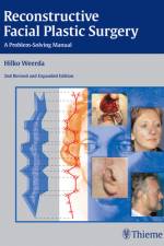
    Drawing on decades of operating room and teaching experience, Dr. Weerda and his team offer a complete guide to reconstructive options for facial, head, and neck defects in this eagerly awaited second edition. Their systematic, step-by-step approach, with an emphasis on meticulous preoperative planning, evaluation of alternatives, and selection of the best procedure, ensures optimal results for all patients. Special features of the second edition: Includes more than 1,500 sequential illustrations of each procedure, along with full-color intraoperative photographs and before and after surgical results Reviews the full range of local, regional, and free flaps used in the reconstruction of facial structures, with chapters on myocutaneous island flaps, delto-pectoral flaps, and free microvascular transplants written by well-known practitioners Offers new and expanded sections on dermabrasion, free flaps, removal of skull bone for modern defect reconstruction, instrument sets/trays, and more Covers the entire scope of the field, from basic principles, anatomy, wound healing, and scar revision to defect closings in each facial region, bone grafts, and the groundbreaking auricular reconstructive techniques developed by Dr. Weerda Focusing on the questions, problems, and technical solutions most commonly encountered in everyday practice, this compact book will be valuable to both the novice and more experienced surgeon. It is filled with the insights, wisdom, and experience of a leading worldwide expert, and will be kept close at hand as a refresher, teaching guide, encyclopedia of facial plastic techniques, and standard operating room reference.

  • von Martin Vahlensieck
    234,00 €

    2. udgave. The value of MR imaging for the evaluation of musculoskeletal system disorders cannot be over-stated. It is the only imaging modality that enables visualization of all components of the joints within single examinations. Yet, given the bewildering variety of possible sequence parameters, with and without contrast medium, acquiring and interpreting MR images with confidence is a challenge, requiring experience usually only gained after examining 1000s of studies with a careful systematic approach. Like the First Edition, the Second Edition of MRI of the Musculoskeletal System assists the radiologist in acquiring the most reliable and complete imaging information, so as to achieve a high degree of diagnostic certainty quickly and efficiently. Key Features: More than 2000 MR images of reference quality, the majority new for this edition Drawings, where helpful, aid the reader in identifying and delineating normal and pathological entities Includes all the latest advanced techniques: MR neurography and myelography, diffusion imaging, quantitative MRI, mDIXON, and more All MR exams described fully, with choice of sequence, positioning, choice of coils, when/how to use contrast, protocols Discussions of possible errors in interpretation Comparison of MR imaging with other modalities Tables expand and organize information on sequence parameters and differential diagnoses More than just an authoritative reference, Vahlensieck's MRI of the Musculoskeletal System is the ideal practical helper to accompany the radiologist at the workstation on a daily basis.

  • von Torsten Bert Moller
    24,00 €

    1. udgave. The key for any beginning radiologist who wishes to recognize pathological findings is to first acquire an ability to distinguish them from normal ones. This outstanding guide gives beginning radiologists the tools they need to systematically approach and recognize normal MR and CT images. Highlights include: Reference-quality images from the author's own teaching files show all standard normal findings as seen in CT and MRI Checklists in each section offer the reader a systematic way to approach the images Thorough guidelines to help beginning radiologists dictate their reports Lists of standard measurements and tips for ruling out pathology.

  • von Mogens Brøndsted Nielsen
    155,00 €

    USB-stick. 1. udgave. Dallas Rhinoplasty and Cosmetic Surgery at your fingertips! The Dallas Rhinoplasty and Dallas Cosmetic Surgery Dissection Video Collection brings the world-renowned Dallas Rhinoplasty and Cosmetic Surgery Courses directly to your computer or mobile device. If you missed the course, you can still dissect with Dr. Rohrich and Dr. Ahmad! Review nasal anatomy and learn the skills and procedures needed to perform a rhinoplasty that produces the best functional and aesthetic result possible. Facial anatomy relevant to face and neck lift surgery, as well as brow lift and blepharoplasty are shown in great detail. This collection offers viewers the following key features: Surgical dissection footage of procedures performed by a legend in plastic surgery and rhinoplasty, Dr. Rod J. Rohrich A 30-minute step-by-step sequence for performing an open rhinoplasty dissection A 30-minute cosmetic surgery demonstration of browlift, blepharoplasty, extended SMAS and Lift and Fill Facelift as well as neck rejuvenation dissection techniques Over an hour of high-quality cadaveric dissection footage with detailed views of the anatomy Clinically relevant procedures for multiple subspecialties by experts in the field A dissection guide with stunning full-color photographs and illustrations of the procedures captured in the video This concise but thorough collection provides the easily accessible, expert content plastic and facial plastic surgeons worldwide have been waiting for from the names they trust most in rhinoplasty and facial rejuvenation surgery.

  • von Matthew M. Hanasono
    94,00 €

    The definitive resource on surgical flaps from renowned MD Anderson plastic surgeonsHandbook of Reconstructive Flaps by Matthew Hanasono and Charles Butler reflects contributions from clinicians associated with the prestigious MD Anderson Cancer Center. The institution is internationally recognized as one of the world's premier reconstructive centers and renowned for its Reconstructive Microsurgery fellowship program. This unique resource provides a comprehensive review of microvascular surgery techniques using the most common reconstructive pedicle flaps, free flaps, perforator flaps, and workhorse flaps.Seven parts organized anatomically detail flaps used for the head/neck, chest, back, abdomen, pelvis, upper extremity, and lower extremity, followed by an eighth part on lymphedema. Each flap or procedure includes pertinent anatomy, indications for usage, preoperative considerations, a concise step-by-step description of the operative setup, the actual procedure, and donor site closure.Key Features:A Pearls and Pitfalls section in each chapter outlines key concepts and critical nuances in surgical techniques or patient managementMore than 200 clear diagrams, line drawings, and illustrative surgical photos enhance understanding of high impact pointsHigh quality videos posted online elucidate flap techniquesA section dedicated to the microsurgical treatment of lymphedema covers lymphovenous bypass, vascularized lymph node transfers from the groin and supraclavicular region, and lymphovenous anastomosisThis is an essential, user-friendly backpack reference for plastic surgery residents to use during rounds and as a board prep. It is also an excellent resource for plastic surgeons new to clinical practice and veteran practitioners as a refresher on unfamiliar flaps.This book includes complimentary access to a digital copy on https://medone.thieme.com.

  • von Richard B. Gunderman
    61,00 €

    Pediatric radiology provides an opportunity to care for perhaps the most vulnerable patients of all, from prenatal life through adolescence. Pediatric Imaging, Second Edition by Richard Gunderman and Lisa Delaney features 100 new cases along with two board-type multiple-choice questions for each. A wide spectrum of cases focusing on radiology in children – from basic to advanced – are strategically designed to increase a resident's knowledge and provide robust exam preparation. For maximum ease of self-assessment, each case begins with the clinical presentation on the right-hand page; study that and then turn the page for imaging findings, differential diagnoses with the definitive diagnosis, essential facts, pearls and pitfalls, and more.

  • von Rebecca Stein-Wexler
    69,00 €

    Top 3 Differentials in Pediatric Radiology by renowned pediatric radiologist Rebecca Stein-Wexler and colleagues is part of a series of radiology case books that mirror the highly acclaimed O'Brien classic, Top 3 Differentials in Radiology. Each case is formatted as a two-page unit. The left page features clinical images, succinctly captioned findings, and pertinent clinical history. The right page includes the key imaging gamut, differential diagnoses rank-ordered by the "Top 3," additional diagnostic considerations, and clinical pearls.This book includes an exceptionally diverse array of common and important gamuts encountered in pediatric radiology practice. It is organized into six sections: head and neck imaging; chest, cardiac, and airway imaging; gastrointestinal imaging; genitourinary imaging; musculoskeletal imaging; and brain and spine imaging. Each section begins with a series of differential-based cases and concludes with important "Roentgen Classics" – cases with imaging findings characteristic of a single diagnosis.

  • von Eugene C. Lin
    86,00 €

    PET and PET/CT have been increasingly used as effective imaging modalities in the management of patients with cancer, neurologic disease, musculoskeletal disease, and cardiac disease. PET and PET/CT: A Clinical Guide, Third Edition by world renowned molecular imaging pioneer Abass Alavi and esteemed diagnostic and nuclear radiologist Eugene Lin features the latest advances in PET technology in an easy-to-read format.The book lays a solid foundation with opening chapters on scanner physics, radionuclide basics, study interpretation, patient preparation, quantitative whole-body PET/CT imaging, normal variants, benign findings, and clinical applications.

  • von Maria Peris-Celda
    230,00 €

    1. udgave. Masterful 2D and 3D head, neck, and brain dissections provide unsurpassed insights into head, neck, and brain anatomy An internationally renowned and beloved author, educator, brain anatomist, and neurosurgeon, Professor Albert Rhoton has a special place in medical history. He was revered by students and colleagues and is regarded as one of the fathers of modern microscopic neurosurgery. A driving principle in his anatomy lab was the simple phrase, "Every Second." This was embraced in his philosophy that every second of every day, a patient's life was improved by a surgeon assisted by the anatomic knowledge his lab helped elucidate and distribute. Rhoton's Atlas of Head, Neck, and Brain is the visually exquisite crowning achievement of Dr. Rhoton's brilliant career and unwavering dedication to the intertwined pursuits of surgical anatomy and neurosurgery. The atlas reflects the unparalleled contributions Dr. Rhoton made to the contemporary understanding of neurosurgical anatomy.

Willkommen bei den Tales Buchfreunden und -freundinnen

Jetzt zum Newsletter anmelden und tolle Angebote und Anregungen für Ihre nächste Lektüre erhalten.